Macular edema is a swelling of the macula, the central part of the retina responsible for sharp, detailed vision — most commonly linked to diabetes but caused by several conditions.
What Is It?
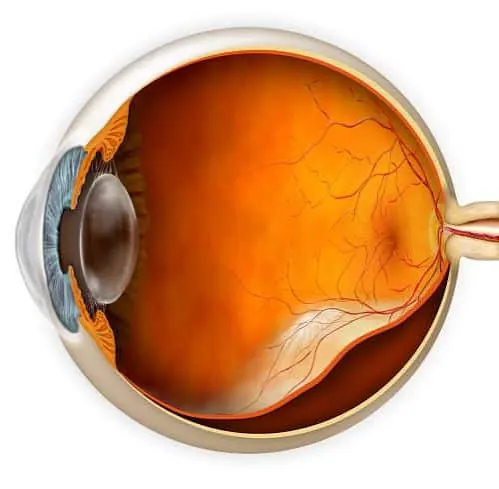
Macular edema occurs when fluid accumulates within the macula, the small central area of the retina responsible for straight-ahead, detailed vision used for reading, recognising faces and driving.
It is most commonly associated with diabetic eye disease (diabetic macular edema), but can also occur after retinal vein occlusion, uveitis (eye inflammation), following certain eye surgeries, or with other retinal conditions.
Because the macula is responsible for central vision, even a small amount of swelling there can noticeably blur or distort vision, even when peripheral (side) vision remains normal.
Symptoms & Indications
Macular edema symptoms are centred on central, detailed vision rather than side vision, and can develop gradually or more suddenly depending on the cause.
Reading, recognising faces or seeing fine detail becomes difficult, while peripheral vision often stays relatively normal.
Straight lines, such as door frames or text, may appear wavy or bent — a sign specifically linked to macular problems.
Some patients notice colours appear duller or less vivid than before.
A blurred or blank patch directly in the centre of vision can occur as swelling progresses.
Treatment Options
Optical Coherence Tomography gives a detailed cross-sectional view of the retina, directly showing the amount and location of fluid in the macula.
Where diabetes, vein occlusion or inflammation is the cause, managing that underlying condition is a key part of treatment.
Depending on the cause and severity, injection therapy or other retinal treatments may be recommended — discussed individually based on OCT findings.
What To Expect
Diagnosing macular edema relies heavily on retinal imaging in addition to a clinical examination.
Pupils are dilated so the macula and retina can be examined in detail.
A quick, non-invasive OCT scan measures retinal thickness and shows fluid accumulation precisely.
Blood sugar status, blood pressure, and any history of eye inflammation or vein occlusion are reviewed to identify the underlying cause.
A treatment and monitoring plan is set based on the cause and severity, with OCT used to track response over time.
Please note: Macular edema can recur, and response to treatment varies between patients and underlying causes. Ongoing monitoring is usually needed even after initial improvement.
Common Questions
Evaluated and treated under Dr. Deepak Gandhi, Director of Gandhi Eye Centre, with clinical experience across cataract, glaucoma, cornea, retina and pediatric eye care.
View Doctor Profile →If you notice blurred, wavy or distorted central vision, book a retinal evaluation at Gandhi Eye Centre, Faridabad.