The retina is the light-sensing layer at the back of your eye. Expert diagnosis and treatment of diabetic retinopathy, retinal detachment, macular degeneration, and vein occlusions — all under one roof in Faridabad.
Retina Procedures
Patient Satisfaction
Years of Excellence
Retina Surgeons
The retina — the light-sensing layer lining the back of the eye
The retina is a thin layer of light-sensitive tissue lining the back of the eye. It converts light into electrical signals sent to the brain via the optic nerve. Damage anywhere in this delicate layer can cause permanent, irreversible vision loss if not treated promptly.
Millions of photoreceptor cells (rods and cones) capture light and convert it into neural signals.
The optic nerve carries these signals from the retina to the brain, forming the images you see.
Diabetes, aging, high myopia, and trauma can damage the retina — causing floaters, distortion, or sudden vision loss.
Modern retina procedures — from injections to microsurgery — can preserve or restore vision when caught early.
Our retina specialists diagnose and treat the full spectrum of retinal conditions with the latest technology available in India.
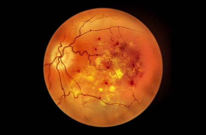
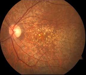
Prolonged high blood sugar damages the tiny blood vessels of the retina, causing bleeding, swelling, and gradual vision loss. India's leading cause of preventable blindness in working-age adults.
The retina peels away from its underlying tissue — a true eye emergency. Sudden flashes, floaters, or a curtain-like shadow demand immediate surgical attention to save vision.
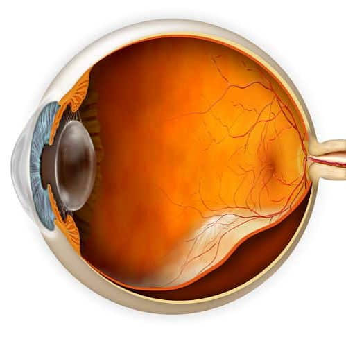
Deterioration of the macula (central retina) causes blurred or distorted central vision, most common in patients over 50. Anti-VEGF injections can slow or halt its progression.
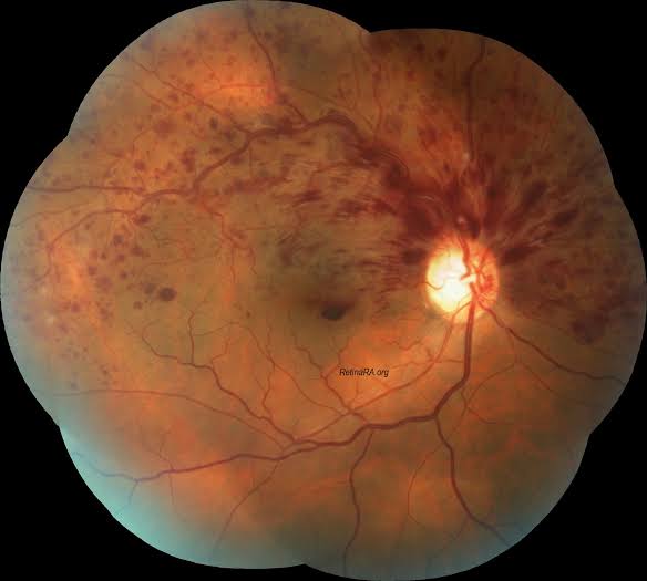
Blockage of a retinal vein causes sudden bleeding and swelling, leading to blurred vision. Often linked to hypertension and needs urgent injection or laser therapy.
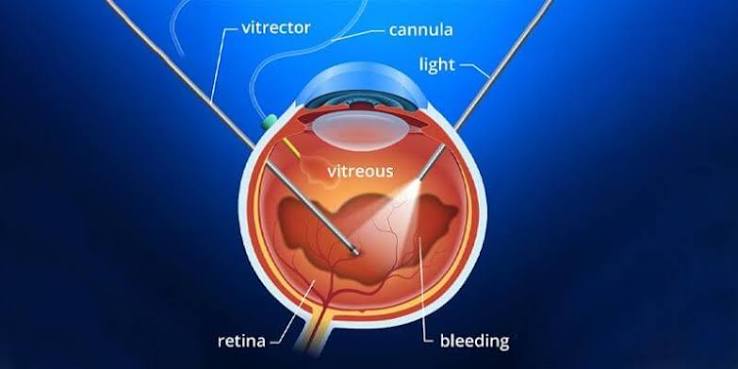
A small break in the macula causes central vision distortion or a blind spot. Vitrectomy surgery can successfully close the hole and restore vision in most cases.
Fluid accumulates under the retina, often triggered by stress or steroid use, causing distorted central vision. Usually managed with observation, laser, or targeted therapy.
Retinal disease often progresses silently until it's advanced — watch out for these warning signs and get examined promptly:
A sudden increase in floating spots or cobweb-like shapes can signal bleeding or a retinal tear.
Brief flashes, especially in peripheral vision, often indicate the retina is being pulled or torn.
A dark curtain or shadow spreading across your field of view is a classic sign of retinal detachment.
Straight lines appearing wavy or bent (metamorphopsia) is a key symptom of macular disease.
Sudden, painless loss of vision in part or all of one eye needs same-day emergency evaluation.
Difficulty reading or recognising faces while peripheral vision stays normal points to macular involvement.
Retinal detachment is an eye emergency. Seek care within hours — every hour of delay reduces the chance of full vision recovery.
Retinal problems arise from systemic disease, aging, genetics, or trauma. Knowing the cause helps guide the right treatment.
Chronically high blood sugar damages retinal blood vessels — the single biggest cause of retina disease in India today. Annual screening is essential for every diabetic.
Uncontrolled high blood pressure damages the delicate retinal blood vessels, raising the risk of vein occlusions and hemorrhages.
Age-related macular degeneration becomes more common after 50, gradually affecting central, detailed vision needed for reading and faces.
Very near-sighted eyes have a thinner, stretched retina that is significantly more prone to tears and detachment.
Certain retinal dystrophies and a family history of AMD or detachment increase your personal risk — early screening is advised.
Blunt or penetrating eye trauma can tear or detach the retina immediately or months after the original injury.
At Gandhi Eye Centre, we use state-of-the-art imaging for precise retinal evaluation — often detecting disease before symptoms appear.
High-resolution photographs of the retina document its current condition and track disease progression over time.
Cross-sectional imaging reveals microscopic retinal layers, detecting fluid, swelling, or holes invisible to the naked eye.
A dye injected into the arm highlights retinal blood vessels, revealing leaks, blockages, or abnormal new vessels.
Used when the retina cannot be viewed directly (e.g. dense cataract or hemorrhage) to check for detachment or masses.
Wide-field examination of the peripheral retina under dilation — essential for detecting tears near the retina's edges.
A dye-free scan that maps retinal blood flow directly, ideal for monitoring diabetic and vascular retinal disease.
Maps your full field of vision to detect peripheral vision loss caused by retinal or optic nerve damage.
A simple grid test that detects even subtle distortion in central vision — useful for monitoring macular disease at home.
From injections to microsurgery — we offer complete retina care under one roof.
Diabetic retinopathy often has no symptoms until vision loss is advanced. Annual screening catches it early, when treatment works best.
Every package includes pre-treatment evaluation and post-treatment follow-up care.
*Prices are indicative. Final cost depends on consultation, severity, and procedure chosen. Insurance support available.
Book a retina examination or get expert advice — our specialist team is here to protect your vision.
Expert retina specialists. Advanced technology. Compassionate care.
Book your retina examination at Gandhi Eye Centre, Faridabad today.